Digital Dermoscopy & Mole Mapping

Digital dermoscopy and mole mapping are modern methods of skin monitoring aimed at the early identification of changes that may require further evaluation. Because significant changes in moles often develop gradually, recording and comparing images over time helps the dermatologist track their progression with greater accuracy.


Two Levels of Examination, a More Comprehensive Picture


Digital Dermoscopy


Digital dermoscopy involves the examination of individual moles using a specialized high-resolution camera with magnification. The image of each lesion is stored digitally so it can be compared during future visits. It is particularly useful when close monitoring of specific moles or initial documentation is required.


Full-Body Mole Mapping


Full-body mapping involves systematic photography of the entire body in standardized positions. This creates a detailed digital record, which allows for image comparison during re-examinations and the identification of new lesions or changes in pre-existing moles.
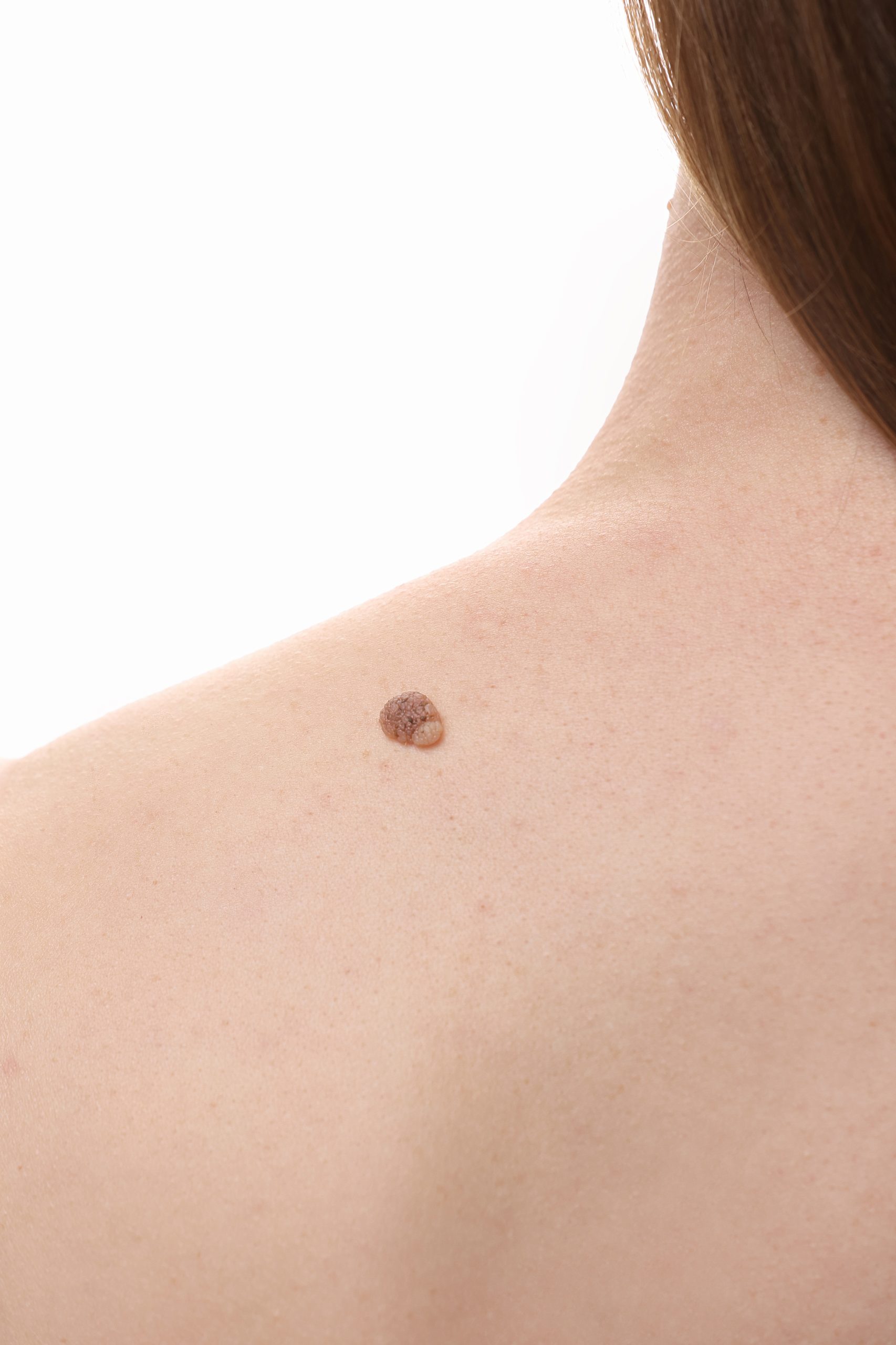
FotoFinder: The System We Use

FotoFinder is a specialized medical system for digital dermoscopy and mole mapping designed for clinical use. The ability to compare images between different visits provides a significant advantage in long-term skin monitoring.


High Resolution and Documentation


Each mole is examined under magnification that helps reveal morphological details not always visible to the naked clinical eye. Images are saved in the patient’s digital file with reference to their exact location on the body.


Image Comparison Over Time


During follow-up appointments, the system allows for the comparison of new and older images, helping the doctor identify potential changes that require closer assessment.


Patient Digital Archive


Each visit is added to the individual’s digital medical history, offering the dermatologist a valuable tool for tracking the evolution of moles over time.


Safe and Painless Procedure


The examination is non-invasive, painless, and does not emit radiation. It requires no special preparation or recovery time.

Digital Dermoscopy vs. Standard Dermoscopy


Includes recording and storing images.

  • Allows comparison with previous visits.
  • Facilitates the monitoring of new lesions or changes.
  • Can also be used for full-body mapping.


Who It Is For


The examination can be particularly beneficial for individuals with:


  • An increased number of moles.
  • Unusual or irregular (atypical) moles.
  • Fair skin phototype or a history of sunburns.
  • A family history of melanoma or other skin cancers.
  • Prior removal of a mole with dysplastic features.
  • Immunosuppression or specific medication that affects the skin.
  • A need for preventive screening with documented follow-up.


Follow-up Frequency


The frequency of follow-up exams is determined by the dermatologist, depending on the individual risk profile and the findings of the initial examination.


  • Every 6–12 months: For individuals at higher risk, such as those with numerous moles, fair skin phototypes, or a family/personal history of melanoma.
  • Every 12 months: For routine annual preventive screenings or lower-risk cases.
  • Every 3–6 months: When closer monitoring is required following the removal of a suspicious lesion or the appearance of a new alteration.

Please enter your phone number and we will contact you shortly

Frequently Asked Questions (FAQs)

How long does the examination take?


Digital dermoscopy of individual moles typically takes 20–30 minutes. Full-body mole mapping can take approximately 45–60 minutes, depending on the number of moles and the extent of the recording.


Is any preparation required?


No special preparation is needed. It is recommended not to apply sunscreen, heavy moisturizer, or body makeup on the day of the exam so that the images are clearer.


What happens if a suspicious mole is detected?


The dermatologist evaluates the finding and, if deemed necessary, may recommend removal for histological examination (biopsy). At Elite Plastic Surgery, the procedure can be organized within the same clinic without unnecessary delays.


Is the examination painful?


No. The procedure is painless, non-invasive, and requires absolutely no downtime.


Can I undergo mole mapping if I have very fair or very dark skin?


Yes. Digital dermoscopy is applicable to all skin phototypes, and the interpretation of the findings is always personalized by the physician.


What is the difference between standard dermoscopy and mole mapping?


Standard dermoscopy evaluates lesions at the exact moment of the examination, whereas digital mole mapping allows for the storage and comparison of images over time. This long-term tracking is its key advantage.